As part of a French collaboration1, BIAM researchers have just demonstrated the decontamination capabilities of a cyanobacterium: Gloeomargarita lithophora, which captures strontium-90 (90Sr), a radionuclide present in power plant effluents. This discovery opens the way to highly effective, environmentally-friendly bioremediation.
Co-discovered several years ago by IMPMC1 in a stromatolite from Lake Alchichica in Mexico, a photosynthetic bacterium is capable of mineralising strontium, an element chemically close to calcium. This particularity caught the attention of Virginie Chapon, head of the IPM team and research director at BIAM, who launched an initial study on the subject: could this cyanobacterium also mineralise strontium-90 (90Sr), a radioelement present in trace amounts in effluents from nuclear power plants? “During this initial study, we were able to demonstrate that Gloeomargarita lithophora trapped 90Sr in the laboratory”, she explains. “However, we still didn’t know whether this bacterium was capable of resisting radioactivity and effectively capturing 90Sr under conditions closer to reality”.
A photosynthetic organism for biological decontamination
The results of a new study carried out in collaboration1 with the I2BC, the IMPMC and the LPCA, as part of the CEA’s FOCUSDEM program, now demonstrate this unambiguously: in the presence of light, G. lithophora captures and stabilises 90Sr for at least 20 days, while surviving radioactivity.
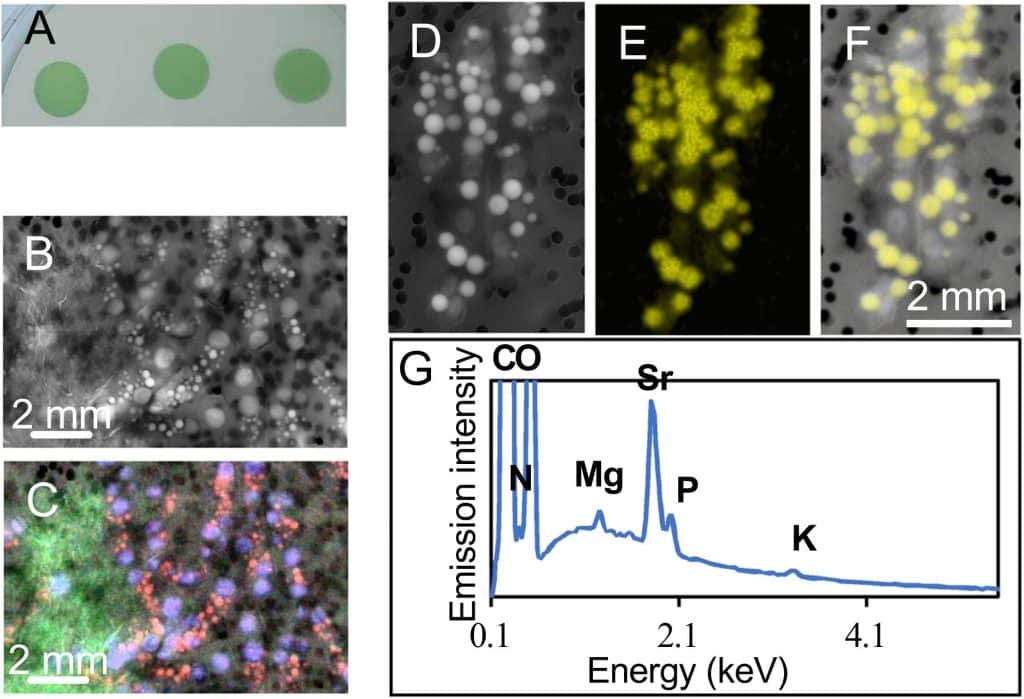
Even more impressive: “This cyanobacterium is capable of storing CO₂ while cleaning up to 98% of an environment simulating real nuclear power plant effluent in just 72 hours. It’s a major scientific breakthrough”, stresses the researcher. Thanks in part to the energy produced by the process of photosynthesis, the bacteria trap the Sr and transform it into granules of minerals visible under the microscope (see visuals).
Equipment at the cutting edge of research
This work once again illustrates the importance of fundamental research in the field of decontamination: “in order to develop biological approaches, we need to study the adaptation of living organisms to environmental constraints in order to understand the mechanisms involved, in particular in this case tolerance to radioactivity and the mecanisms of 90Sr sequestration. This is a necessary step before envisaging a move to industrial scale‘, she points out, before emphasising that ’BIAM2‘s unique infrastructure has enabled us to study photosynthetic micro-organisms in the presence of radioactive elements, a sine qua non for carrying out these experiments”.
Outlook
This study opens up promising avenues, both fundamental and applied. The next steps will provide a better understanding of the tolerance of G. lithophora to radioactivity, as well as the mechanisms of transport and sequestration of 90Sr. G. lithophora now presents an innovative alternative to conventional decontamination methods (chemical precipitation or adsorption on solid materials), offering a biological alternative for the nuclear industry.

Growth and formation of intracellular calcium and strontium carbonate granules by the nearly axenic Gloeomargarita lithophora.
A) Growth of G. lithophora spotted on MM agar plate with 2mM thiosulfate after axenisation. B) SEM images in the secondary electron (SE) mode of the cells from almost axenic G. lithophora grown in MM allow detection of iACC (smaller brightest spots) together with polyphosphate granules (large bright grey). C) Overlay of Ca (red), P (blue) and Si (green) chemical maps obtained by energy dispersive X-ray spectrometry over the AsB SEM image (grey levels) shown in (B). D) SEM images in the secondary electron (SE) mode of the cells from almost axenic G. lithophora grown in the presence of 10 mM Sr in the mineral medium. E) Sr (yellow) chemical map obtained by EDXS F) Overlay of Ca (red) and P (bleu) Sr (yellow) chemical maps obtained by EDXS and corrected from the bremsshtrahlung over the AsB SEM image (grey level) shown in (D). G) Average EDXS spectrum of the area scanned in D.
1This study was carried out in collaboration with the Institut de Biologie Intégrative de la Cellule – CNRS (I2BC, Saclay), the Institut de minéralogie, de physique des matériaux et de Cosmochimie UMR 7590 – Sorbonne Université/CNRS/MNHN/IRD (IMPMC, Paris) and the Laboratoire de Physique de Clermont Auvergne (LPCA, Clermont-Ferrand) and as part of the CEA’s FOCUSDEM program, which funded a thesis at BIAM. (link to each institute and laboratory).
2BIAM has a laboratory specifically equipped to carry out microbiology experiments on photosynthetic organisms in the presence of radioactive elements, which is unique in the French research landscape.
Authors :
References